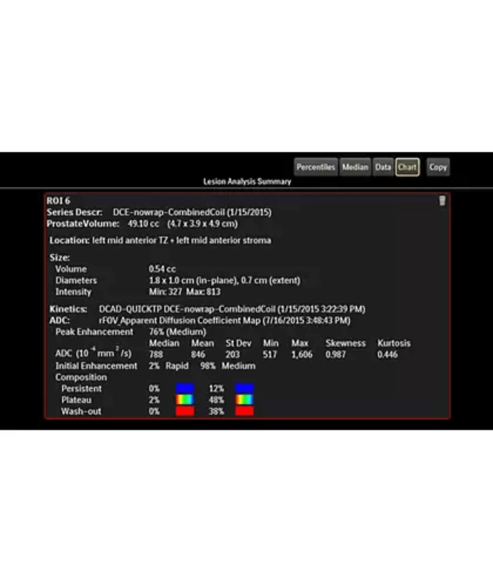
Lesion segmentation enhances workflow efficiency
Launch an automatic segmentation feature with one mouse click. The advanced segmentation algorithm allows for your on-the-fly modification and presents you with a volume analysis, lesion composition statistics, histograms, and a 3D-rendered morphological overview. Auto-populated regions of interest (ROIs) can be viewed as a 2D image or can be mapped onto a maximum intensity projection, where it will appear as a 3D object. The resulting segmentation report provides a calculation of lesion location diameter measurements and location.
Automated reports capture and share relevant data
Use the structured reporting system in DynaCAD Breast to produce highly customized exam reports. You can program reports to automatically append pre-selected images containing kinetic data, measurements, and annotations. The system also automatically populates additional report fields such as lesion diameter measurements, lesion to landmark distances, and volumetric data. Upon completion, you can print patient reports, save as a PDF, or send as DICOM images.


