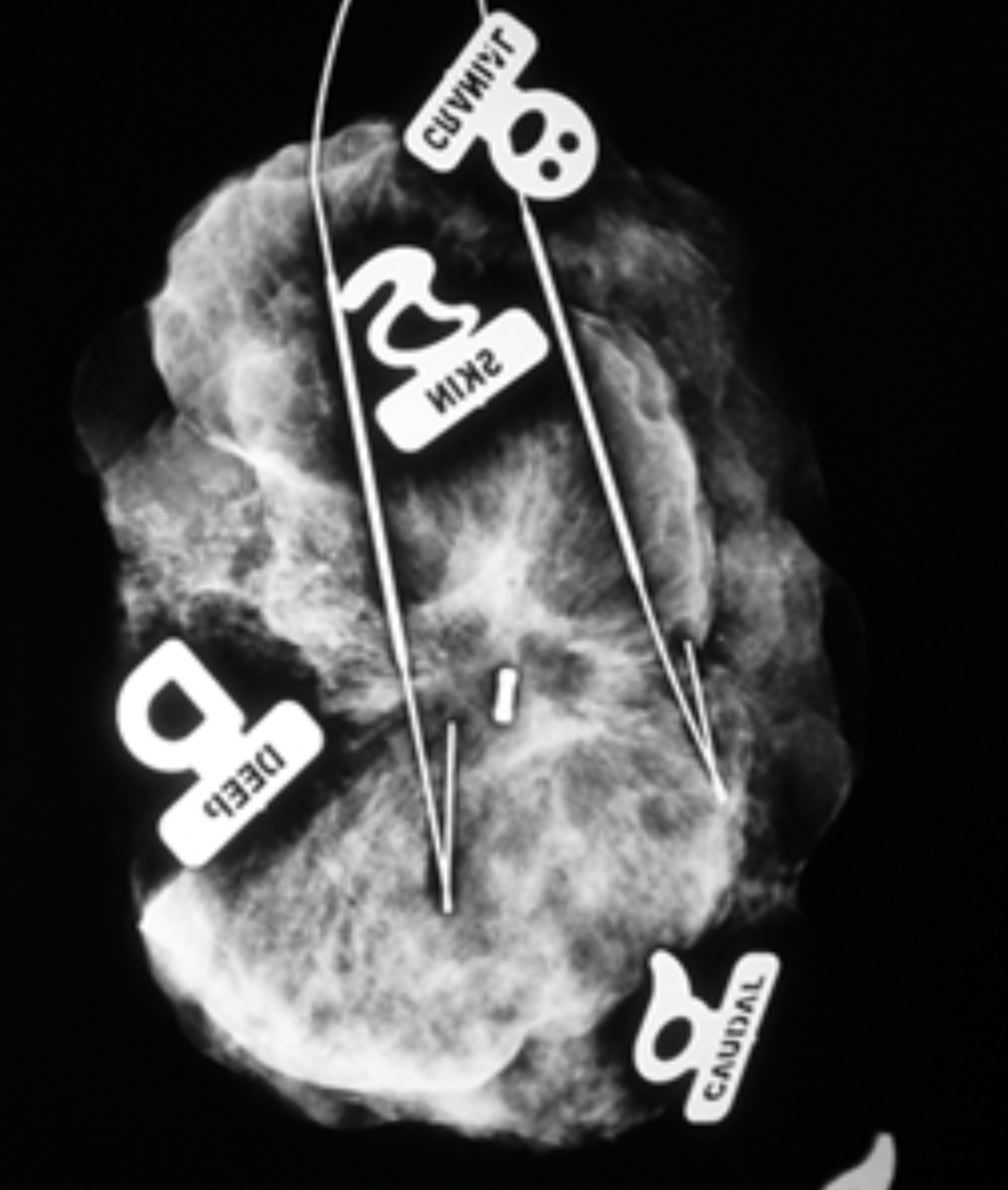
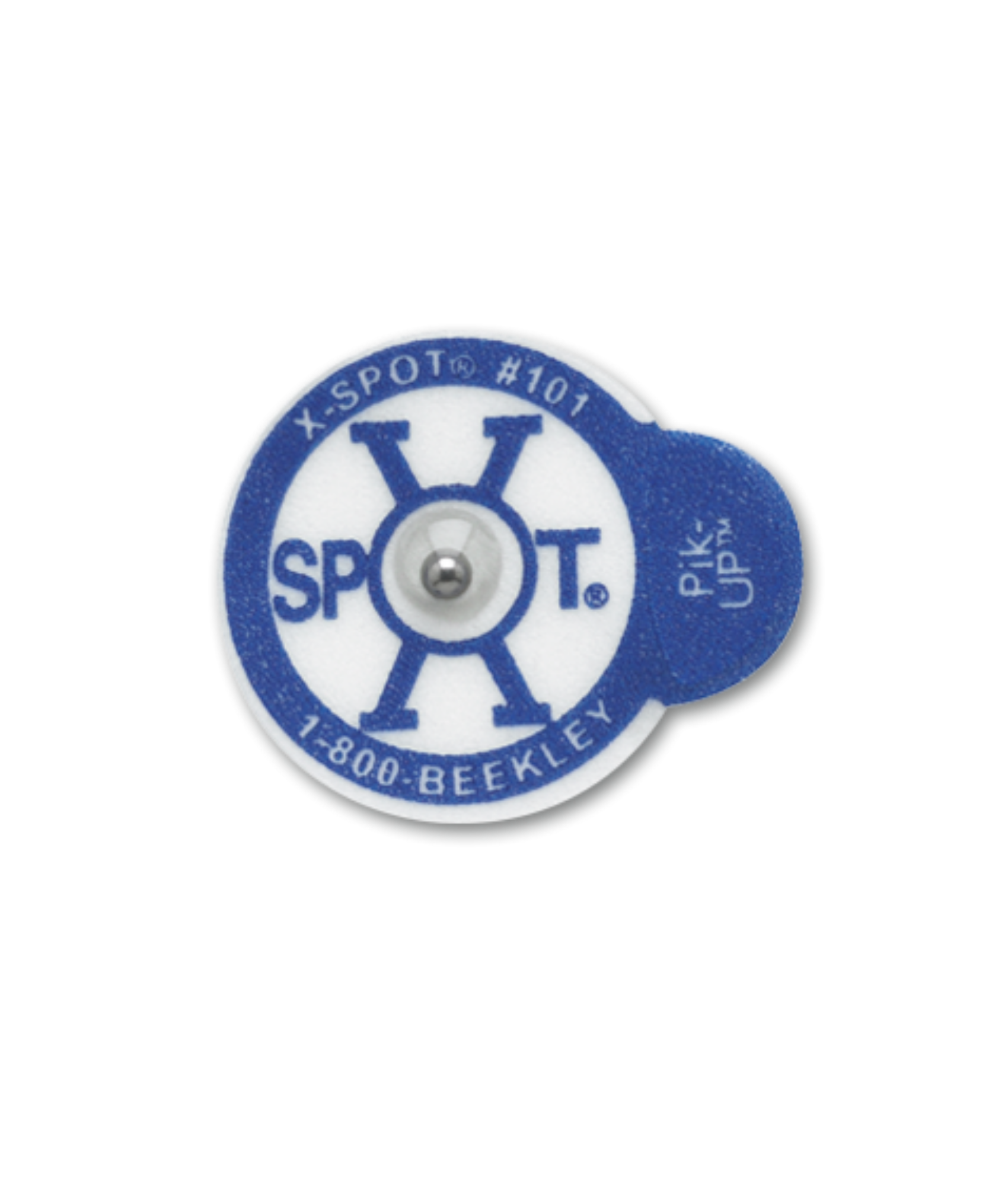
TomoSPOT skin markers are tested and proven to communicate with minimal distraction in digital breast tomosynthesis, or 3D mammography.
Digital Breast Tomosynthesis reveals greater tissue detail in high resolution slices, increasing the number of images a radiologist must review.
Despite its enhanced imaging, questions and uncertainties regarding findings still arise in 3D mammography
That area of architectural distortion could be from a previous surgery, or it could be something new. That suspicious superficial lesion could be a skin mole, or it could be a cancer.
TomoSPOT’s five distinct shapes subtly, yet clearly, communicate the location of moles, skin scars from prior surgeries, palpable masses, nipple position, focal pain or other non-palpable areas of concern with clear visualization of underlying tissue directly on the mammographic image.
A proactive, routine skin marking protocol in 3D mammography with TomoSPOT has been proven to improve specificity and:
- Reduce risk of false negatives and false positives
- Reduce unnecessary additional views and callbacks
- Provide visual documentation of breast anatomy from year to year and when transferring images
- Reduce radiologist reading time on average by 1.34 minutes per case¹
1Tomosynthesis Abstract Preview Radiologist Read-Time


