Description
MRI-guided In-bore Prostate Biopsy, What to Expect?
For the biopsy, the patient will be in prone position on a cushioned table in the MRI scanner. An MRI compatible needle guide will be gently placed into the rectum and connected to the RCM. A combination of MR images and dedicated software will be utilized to position the needle guide for optimal targeting of the prostate lesion and take the biopsy.
System Overview
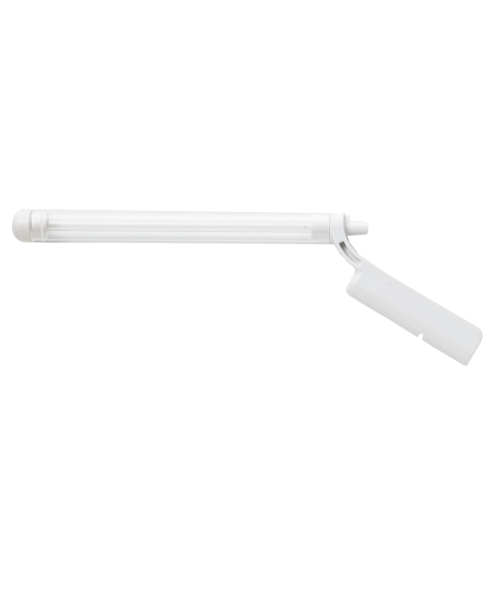
The main component of the system consist of a fully MR compatible manipulator made of high quality plastics. This manipulator is connected via 7-8 meters of tubing to a control unit using a wall feed. The control unit is based in the MR control room and relays the motion derived from the dedicated software system.
Wall feed: Requires 90mm diameter wave guide
Easy Storage
The RCM system typically will be delivered on a cart. This makes the system easy to store and setup for the procedure. It also helps to minimize the clutter in the control room when the system is not used.
Besides the manipulator the cart stores the control unit, air supply via a compressor and optional components like computer/laptop. A long tube connects the control unit with the manipulator inside the MRI room, ideally through a wave guide.
Dimensions: 1145mm x 710mm x 620mm (HxWxD)
Weight: approx. 90kg
Workflow
With the easy-to-learn workflow the software guides the user through the steps of performing the biopsy. Based on an intuitive tab system the received images are used to reidentify the target area. After the calibration of the device relative to the patient it allows the user to select the target on the MR image and move the needle guide towards this position. A fast scanning sequence confirms the location and enables the physician to take the biopsy sample.
Due to the flexibility of the placement of the RCM on the table plate the users can focus on the positioning of the patient on the scanner table. With the preparation and relative comfortable patient positioning, the procedure can be executed in the least amount of time.
Soteria provides online demonstrations of the Remote Controlled Manipulator for MR-guided prostate biopsies on request. During this interactive demonstration, you will have the opportunity to ask questions to our colleagues and to learn more about the opportunities for your institute.
Prostate Diagnosis
MRI In-bore Biopsy
An MR-guided in-bore biopsy is an advanced medical procedure used to collect targeted tissue samples from the prostate gland for the examination of prostate cancer. This procedure employs the high sensitivity of MRI imaging, specialized equipment and software to plan and guide the biopsy route. This ensures that the sample is taken at the exact location of the suspicious area
on the MRI, resulting in highly accurate results.
The MR-guided in-bore biopsy represents a remarkable advancement in prostate cancer diagnosis by significantly reducing uncertainties and errors in the process. In-bore biopsy is typically used to collect 2-3 samples of tumor suspicious areas only and thereby lowering the number of overall cores and complications. The real-time MRI feedback of the needle guide location provides high confidence of the biopsy results and true aggressiveness of potential prostate cancer.
Traditional biopsy methods often had limitations in terms of precision and the ability to accurately locate and target suspicious lesions. With Soteria’s innovative approach, physicians are enabled to detect prostate cancer earlier and treat more precisely. It is a significant step forward in the battle against
this common form of cancer, improving patients’ prognosis and optimizing treatment options.
Benefits of In-bore Biopsy
- High precision: In-bore biopsy offers exceptional precision and accuracy in targeting suspicious areas within the prostate, reducing the likelihood of false-negative results.
- Increased confidence: In-bore biopsy minimizes the risk of sampling errors, ensuring that biopsy cores are taken from the exact tumor suspicious areas.
- Tailored treatment plans: The precise information obtained from in-bore biopsies helps in the development of personalized treatment plans based on the location and aggressiveness of the cancer.
In-bore biopsy is considered a valuable tool in the diagnosis and risk assessment of prostate cancer, especially when highly detailed imaging and precise targeting of suspicious areas are required by combining high sensitivity with high specificity. It offers significant advantages in terms of accuracy and helps to guide treatment decisions for patients.
Get the Optimal Diagnosis
MRI-guided prostate biopsies are essential for the diagnosis and treatment of prostate cancer. Utilizing the precision and detailed imaging capabilities of MRI, in-bore biopsies provide critical information for identifying, targeting and sampling suspicious areas within the prostate gland.
The Revolutionary New Way of Taking Prostate Biopsies
The advanced technique of the Remote Controlled Manipulator significantly enhances diagnostic accuracy. It reduces the need for unnecessary biopsies, minimizes patient discomfort and improves personalized treatment decisions. Ultimately, this approach leads to improved patient outcomes and more effective management of prostate cancer.


